Keratoconus
Keratoconus is a progressive eye disorder that can lead to severe visual impairment if left untreated. Keratoconus treatment includes spectacles and contact lenses for mild to moderate cases and keratoconus surgery (cross-linkage or keratoplasty for more advanced or progressive cases).
While keratoconus is less common than other eye conditions, it still affects a significant number of people. It’s estimated to affect around 1 in 2,000 people – although some studies say it could be more.
It’s important to have regular eye exams, especially if there’s a family history of keratoconus, because the earliest signs may be imperceptible and catching it at an early stage can impact the long-term quality of your vision.
What is Keratoconus?
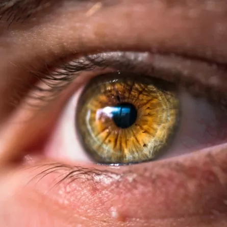
Keratoconus is a progressive eye disorder that causes the normally round cornea – the clear front surface of the eye – to thin and bulge, like a cone.. Everyday tasks such as reading or driving become far more challenging. Since the cornea’s responsibility is bending light into the eye, any change in its shape creates a blurry, distorted image, often requiring specialist contact lenses or other interventions to compensate.
Aetiology of keratoconus is multifactorial with genetic and environmental factors playing an important role. Some people have a family history of the disease, or a history of atopy/allergies or genetic conditions such as downs syndrome. Further pre-disposing risk factors include environmental factors such as excessive eye rubbing
The Symptoms of keratoconus
Keratoconus symptoms can begin with mild vision changes (blurred or distorted sight) and may progress to increased light and glare sensitivity and a rapid increase in short-sightedness and astigmatism . If it’s worse in one eye than the other, you might experience double vision or ghost images, especially when you look at lights.
Causes of keratoconus
In the past, and even still today, the causes of keratoconus were not known. Some recent research has proposed that the corneal tissue that loses strength because of the disease can be caused by an imbalance of enzymes in the cornea.
It is estimated to affect around one in 2,000 people and is equally prevalent in men and women. It’s also more common in some ethnic groups. For example, in people of South Asian origin, it affects around one in 450.
Keratoconus appears to have a hereditary link, asten to 14 per cent of patients with keratoconus have a family history of this eye condition.
As the cornea’s shape changes, vision can shift and become very poor. In the most advanced stages of keratoconus, scarring can result in the loss of transparency of the cornea (corneal hydrops) and the eye may be unable to focus.
Can Keratoconus be Cured?
Keratoconus is often detected when assessing other eye conditions.
The main treatment available for keratoconus is to correct the vision problems that are caused by the irregular cornea. This often means using glasses or soft contact lenses to correct the problems. As the condition worsens though, vision will only be able to be corrected with hard contact lenses that are rigid gas permeable However, this only treats the symptoms of the disorder, and not the underlying cause.
Where the cornea has already thinned this is unfortunately irreversible however a technique of corneal cross-linking surgery is used to strengthen the tissue in keratoconus eyes halting the condition.
Treatments for Keratoconus
For mild cases of keratoconus spectacle lenses can correct the eye shape for shorted sightedness and astigmatism. As the condition progresses to worse than driving standards and can no longer be corrected by spectacles specialist Rigid Gas Permeable lenses are used to correct the cone shape. For progressive keratoconus early referral by your eye health care provider (optician or optometrist) is important, corneal cross-linking treatment can strengthen the cornea and halt the condition helping to preserve vision.
In advanced cases (up to 20% of cases of keratoconus) corneal transplantation (keratoplasty) may be required to restore clearer vision.
