
Diane Regan - "Seen quickly and treatment carried out almost immediately, very efficient."
4.8 / 5 Rated excellent on Trustpilot
14 Top Rated Private Hospitals across the country
Detached Retina (Retinal Detachment)
Retinal detachment is a serious but highly treatable condition that demands swift clinical action to protect your sight from permanent damage. Maintaining clear, uninterrupted vision is vital to your everyday life, which is why sudden visual disturbances require immediate, expert attention.
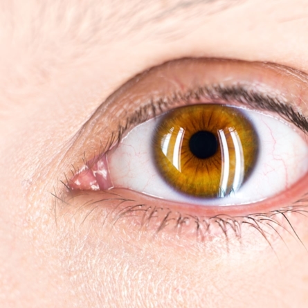
What is a Detached Retina?
A detached retina is a sight-threatening medical emergency that occurs when the light-sensitive layer at the back of the eye pulls away from the vital blood vessels that keep it healthy. The process often starts as a partially detached retina, where only a small, localised section has come loose. However, a partial separation is highly unstable and will rapidly progress to a full detachment, risking permanent blindness if no immediate specialist treatment is provided.
What Causes a Detached Retina?
The natural ageing process of the eye is the primary cause of a detached retina. While age-related changes are the most common trigger, a detached retina can be caused by a variety of underlying structural strains, injuries, and medical conditions:
- Posterior Vitreous Detachment (PVD): This natural, age-related shrinking of the eye’s vitreous gel can tug on the delicate retina, causing a physical tear.
- Severe Short-Sightedness (Myopia): Highly short-sighted eyes are often physically longer, which stretches the retina and makes the tissue thinner, more fragile, and prone to tearing.
- Past Eye Surgery: Previous ocular procedures, most notably cataract surgery, can alter the internal dynamics of the eye and increase the long-term risk of a tear.
- Diabetic Retinopathy: Advanced diabetes can lead to the growth of scar tissue on the retinal surface; as this tissue contracts, it physically pulls the retina away from the back of the eye.
- Trauma or Injury: A severe blow to the face, head, or directly to the eye can cause sudden internal structural forces that instantly tear or detach the retinal tissue.
- Family History: An inherited genetic predisposition to thin retinal tissue or a family history of retinal issues significantly elevates your baseline risk profile.
Recognising Detached Retina Symptoms and Signs
Time is the most critical variable when managing a retinal event. Early recognition of the symptoms of a detached retina is essential for preserving your sight. Because the retina does not possess any pain receptors, a tear or detachment is completely painless, so you must keep a close eye on subtle visual anomalies to identify a potential issue.
The earliest clinical signs of a detached retina involve a sudden onset of floaters, typically dark spots, threads, or cobwebs drifting across your field of vision. This symptom is frequently accompanied by flashes of light in your peripheral vision, caused by the physical mechanical pulling of the vitreous gel against the sensitive neural tissue. As the detachment spreads, you may notice a blind spot in your vision with many patients reporting a dark curtain veil or shadow in their vision. Experiencing any of these symptoms could be a sign of detached retina, and you must seek medical help immediately.
Can an Eye Test Detect a Detached Retina?
Yes, a thorough eye test can detect a detached retina, you can expect your pupils to be dilated if reporting signs or symptoms of a detachment includes pupil dilation. Dilation allows an optometrist to use specialised equipment to inspect the furthest edges of the retina where tears and separations typically begin. However, if you’re actively experiencing emergency symptoms like flashing lights or a dark shadow across your vision, you should bypass a routine high-street optician appointment entirely. Instead, you must seek an immediate assessment at a dedicated eye casualty department, specialist ophthalmic clinic or your local accident and emergency for urgent diagnostic imaging.
Vitrectomy for Cataract Surgery Complications
A vitrectomy is an advanced surgical procedure used to treat rare but serious cataract surgery complications, such as a retinal tear or detachment. Vitrectomy is the surgical removal of the vitreous. Gas is then used to fill the vitreous cavity and to push the detached retina back against the eye wall. The area around the hole is sealed using either laser or cryotherapy to form a permanent scar. The gas bubble reabsorbs spontaneously, and the scarring prevents re-detachment. This is usually carried out under local anaesthesia.
Scleral buckling
Tiny silicone bands or sponge material indent the eye wall, pushing it in towards the detaching retina. Help to reduce the amount that the vitreous gel pulls on the retina (vitreous traction).
A vitrectomy may also be necessary, followed by laser or cryotherapy to produce a permanent seal around the retinal tears.
Can You Drive with a Detached Retina?
No, you must not drive with a detached retina under any circumstances. The sudden loss of peripheral awareness and the compromise of your central visual field make driving exceptionally hazardous and illegal. You must cease driving immediately upon the onset of symptoms or diagnosis. You cannot return to the wheel until your consultant explicitly confirms that your post-surgery vision meets the legal eyesight standards required by the DVLA.
Can You Fly with a Detached Retina?
No, you cannot fly with a detached retina, and you must avoid air travel entirely if you are awaiting treatment or recovering from specific surgical repairs. Flying with an untreated retinal tear or detachment is dangerous because cabin pressure fluctuations can cause the tissue to separate further.
