Laser Eye Surgery
From Only
£25/mo
4.8 / 5 Rated excellent on Trustpilot
15 Top Rated Private Hospitals across the country

From Only
£25/mo

Daily appointments available from the convenience of your own home.
Private Treatments Only.
NHS queries 0207 509 4186
Daily appointments available from the convenience of your own home.
Private Treatments Only.
NHS queries 0207 509 4186


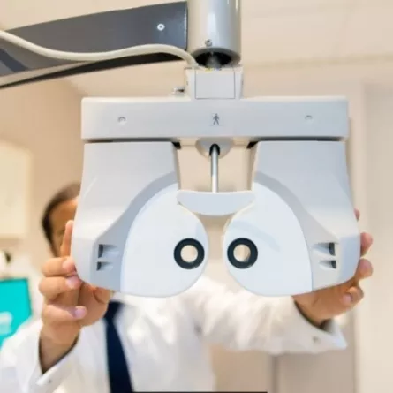
Laser eye surgery is a procedure that corrects blurry vision caused by refractive error, to give you a life without glasses or contact lenses. Laser eye surgery can specifically correct refractive conditions such as short-sightedness, long-sightedness, and astigmatism.
There are three main types of laser eye surgery; SMILE, LASIK and LASEK. For patients undergoing SMILE and LASIK eye surgery, the procedure is quick and painless and there is minimal downtime for recovery post-operatively. For patients undergoing LASEK eye surgery, the procedure is also quick and painless, but you can expect some discomfort which should settle within a week or so.
Our specialist laser eye surgeons are some of the best in the industry, so you’ll feel confident during the process in their experience and expertise.
If laser eye surgery isn’t for you, Implantable Contact Lens (ICL) surgery is an alternative solution.
Laser vision correction works by correcting the focus of the cornea. The different types of refractive eye surgery are: LASIK, LASEK, ReLEx SMILE, PRESBYOND. We are the only UK group to offer all these options, based on what is ideal for your unique vision correction needs.
At Optegra, we offer a range of advanced laser eye surgery procedures designed to gently reshape the cornea and correct vision problems such as short sight, long sight and astigmatism. From the moment you arrive at one of our specialist hospitals,, our expert clinical team will ensure you feel comfortable, informed and fully supported.
Your surgeon will recommend the most suitable treatment based on your eye health, prescription and lifestyle. The most common procedures include:
LASIK
A femtosecond laser is used to create a thin flap on the surface of the cornea. This flap is carefully lifted so an excimer laser can reshape the underlying tissue, correcting your vision. The flap is then repositioned, where it heals naturally without stitches.
LASEK
Rather than creating a flap, the outer layer of the cornea is gently loosened and moved aside. The corneal surface is then reshaped using an excimer laser before the outer layer is returned to its position. A protective contact lens is placed over the eye to support healing.
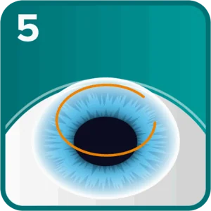
ReLEx SMILE
This minimally invasive procedure reshapes the cornea without creating a flap. A laser forms a small piece of tissue within the cornea, which is removed through a tiny opening. This allows for faster healing and a comfortable recovery.
PRESBYOND®
PRESBYOND® uses advanced wavefront laser technology to improve vision at near, indicating and distance ranges. Each eye is treated slightly differently to provide a smooth range of focus, reducing reliance on glasses. The procedure is quick and painless, with most patients returning to normal activities within a few days.
Your consultant will talk you through each option in detail and help you choose the procedure that best suits your vision needs and lifestyle.
*Diagram specific to LASIK and Presbyond
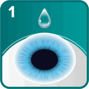
Anaesthetic eye drops applied to ensure a pain-free procedure
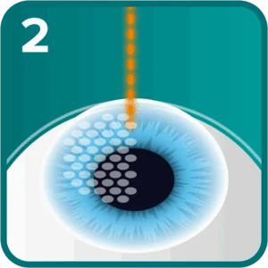
A flap is created in the cornea using a femtosecond laser
The flap is gently lifted to expose the corneal bed
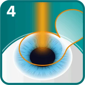
Within seconds the cornea is reshaped by an excimer laser
The flap is gently repositioned by the expert surgeon
During your free initial consultation, your consultant will assess your eyes and recommend the most suitable treatment based on your individual needs. We believe in complete transparency, which is why we offer a clear, single pricing structure for our laser eye surgery procedures.
To make treatment more accessible, we also provide a range of flexible payment options, including up to 24 months interest free. Our indicative price range is outlined below to help you plan with confidence.
Best suited for people with thin corneas and low-moderate prescriptions.
Can treat a wide range of prescriptions and popular for a fast recovery.
The most advanced method with fast recovery using well established keyhole technology.
Blended vision is ideal for age related near changes and helps people see far-middle-near.
Important Information:
Complex Laser is charged at £2,895 per eye. Interest free credit & finance options available.
Learn more about the cost of laser eye surgery.

Laser eye surgery is ideal if you are affected by short sight, long sight or astigmatism. Short-sightedness can range from mild, where treatment may not be required, to severe, where objects will appear blurry.
In the long term, long-sightedness can significantly cause headaches and tiredness due to “over-focus” and lead to double vision. Depending on the seriousness of the problem contact lenses or glasses can be worn but some people may find it less of a hassle to have laser eye surgery.
Laser eye surgery is one of the safest and most well-established vision correction procedures available today. While every surgical treatment carries a small level of risk, millions of people worldwide have safely undergone laser eye surgery, with excellent outcomes. At Optegra, all procedures are performed by highly experienced consultant surgeons using advanced laser technology in purpose-built clinical environments.
For most patients, laser eye surgery offers long-lasting vision improvement with minimal disruption to daily life. Benefits include:
Your consultant will carry out a thorough assessment to ensure laser eye surgery is safe and suitable for you, and will talk you through any risks or considerations so you can make an informed decision with confidence.
Optegra laser eye surgery is very safe and effective. Over 99% of our customers achieved 20/20 vision or better on the next day after the treatment although it may take slightly longer for some patients.
Our world class laser eye consultants are NHS-trained, as well as being Fellows of the Royal College of Ophthalmologists or equivalent organisations. Our ophthalmic surgeons are the best in the industry so you can rest assured of their experience and expertise.

Aftercare is an extremely important part of our laser eye surgery treatment. Our well-rounded aftercare programme is designed to ensure that every patient gets back on their feet in no time at all. It starts right after the day of your laser eye surgery and is tailored to your individual needs. We will regularly check on your recovery to ensure that you get back on your feet in no time.
All Optegra patients have access to the professional advice of our team of fully NHS trained surgeons and staff to answer any of your questions on eye health or procedure.
We have purpose-built Optegra hospitals across England that provide laser eye surgery. Find your nearest hospital below:
As well as conveniently located outreach eye clinics in:
Laser eye surgery takes 15 minutes to perform, but the actual laser takes a very small part of this time. However, we would advise that you allocate 2-3 hours during the day for a correct preparation and initial aftercare steps to take place. At Optegra, we have invested in state-of-the-art advanced laser eye technology to ensure the best possible results for our patients, meaning our patients’ experience is quicker and more comfortable.
Most patients will see the results of laser eye surgery 1-2 days after their surgery. Since everyone is different, some patients enjoy a rapid visual recovery after laser eye surgery and can notice improvements in their vision immediately.
After a patient has had laser eye surgery, they can usually return to work within 24 hours after LASIK or SMILE laser eye treatment, and within a few days after LASEK laser eye treatment. However, every individual is different and it is important that all patients take as much time as they need to recover. Our laser eye surgeons, or a member of our team, will be on-hand to provide any advice or reassurance during the recovery period. In general, most patients are able to go back to work and drive after 48 hours.
Effectively, by reshaping the cornea, laser eye surgery will permanently correct a patient’s vision. LASIK, LASEK and SMILE Laser eye surgery treatments are considered a permanent cure for most refractive errors, however some patients may find they need reading glasses at a point in their lives as the eye naturally ages. This condition is also known as presbyopia.
While many people are apprehensive about pain during their laser eye surgery procedure, in reality, it is comfortable and pain-free. There is also very little discomfort felt following the procedure, so there is nothing to worry about if fear of pain is stopping you from having surgery.
Laser eye surgery is typically a quick procedure. The actual time will vary depending on the type of laser procedure you have opted for. SMILE and LASIK typically take 15 minutes per eye, while LASEK takes around 20 minutes per eye.
Yes. Our specialist Monovision technique can help correct reading vision difficulties caused by presbyopia. However, LASIK, LASEK or SMILE surgeries may not always be suitable for people who need strong prescriptions to read. In this case an alternative treatment may be suggested.
If you are unsure whether you are suitable for laser eye surgery, please don’t hesitate to get in touch to arrange a free, no-obligation consultation with one of our laser eye surgeons, and see how we can help you.
In short, the answer is yes. However, each case is unique, so your consultant will assess individual suitability to ensure that you are eligible for another treatment. Essentially, it will depend on the amount of tissue in the eye and how much of it was removed during the first surgery.
At Optegra, we do not perform laser eye surgery on anyone aged below 21. This is because the eyes are still developing during childhood and adolescence, and vision changes can occur during this period.
For older patients, it is not recommended to have laser eye surgery past the age of 40, as they might experience age-related vision changes imminently requiring them to wear glasses. For patients who are around 40, Presbyond is a unique laser treatment that could be more suitable.
Yes. In fact, laser eye surgery is a highly effective method for correcting astigmatism, along with other refractive errors like nearsightedness (myopia) and farsightedness (hyperopia).

Not ready for a consultation? Learn more about our range of treatments, doctors and hospitals
Information pack
Book your virtual consultation with our top rated eye hospitals
Book Now
We'll answer any questions you may have about treatment.
Private: Mon-Thu: 8am – 6pm, Fri: 8am – 5.30pm NHS: Mon-Fri: 8am – 6pm

Manage your existing bookings & payments
Patient Portal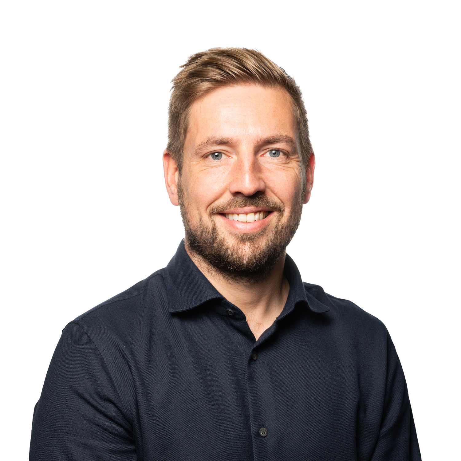
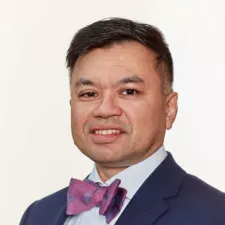
Dr Alastair Stuart is Medical Director at Optegra, and has extensive experience in both Laser eye surgery and Cataract Surgery.
Medically Reviewed Date: 12th June 2026